


Unveiling the structure of the human cornea
This text is a translation of the text published in French on the CNRS website.
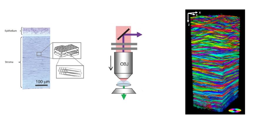
One of the most important functions of the cornea is to help focus light onto our retina. Its curved shape, and therefore its ability to focus light, must be perfectly stable to ensure a clear image despite the various shocks and rubs received over the course of a day. The cornea is therefore composed of several hundred collagen lamellae stacked one on top of the other in various orientations. However, the transparency of this tissue, essential for visual acuity, hinders its imaging by conventional optical microscopes. In fact, the poor characterization of the lamellar structure of the cornea today limits our understanding of the relationship between its structure and function, particularly with respect to its mechanical properties. As a result, certain pathologies associated with defective corneal structure, such as keratoconus, remain poorly understood.
In a recent work, a team from the Optics and Biosciences Laboratory used a recent three-dimensional optical imaging technique, second harmonic generation microscopy (known as "SHG" microscopy), to specifically visualize collagen without any prior labeling. In a unique way, their approach takes advantage of the light polarization (the direction of the electric field of the optical wave) to reveal the direction of the collagen fibrils that make up the lamellae of the cornea. This imaging technique is implemented by detecting the reflected signal, which means it could eventually be used for in vivo diagnosis, and also gives far more accurate results than with the transmitted signal. Ten intact human corneas were characterized and used to validate the method, since SHG polarimetry had never been used on tissue of such thickness (around 600 µm). In particular, the researchers demonstrated for the first time that, while the lamellae of the human cornea are globally oriented in two perpendicular directions, their predominant orientation changes with depth.
This study opens the way to promising new characterizations of the cornea, such as mapping the size and distribution of lamellae as a function of depth, but also as a function of position (center or periphery of the tissue). This information will feed into mechanical modelling of corneal behaviour during variations in intraocular pressure or healing processes. Finally, the study of pathological tissues will clarify the role of the corneal defective structure in certain diseases.