


Diagnosis using polarized light
A pregnant woman arrives at the emergency room with symptoms of premature labor. In order to make a diagnosis, the practitioner measures the length of the cervix using a transvaginal ultrasound. Below a certain threshold, he considers the risk to be very real. The expectant mother is then hospitalized and given treatment to reduce her uterine contractions. However, there are no recommendations on the threshold to be taken into account to detect a risk of prematurity, and it is currently impossible to establish a precise correlation between cervical length and the timing of delivery. “The lack of a reliable diagnostic technique makes premature birth the leading cause of perinatal mortality worldwide. In addition, many pregnant women are hospitalized unnecessarily and exposed to the side effects of inappropriate treatments, which represents a considerable healthcare cost,” explains Angelo Pierangelo.
At the Laboratory of Physics of Interfaces and Thin Films (LPICM*) at Ecole Polytechnique, the researcher and his team are exploring the physical properties of light—specifically polarization—to diagnose premature births more reliably. According to the theory of electromagnetism, light consists of a strongly interconnected electric and magnetic field that oscillates in space and time. Polarization is therefore the space-time trajectory of the electric field. “When polarized light interacts with tissue, its polarization changes according to the microscopic properties of the target,” explains the researcher. He then uses a specific imaging technique—Mueller polarimetric imaging—to obtain a complete characterization of the polarimetric properties of biological tissue.
Exploring tissues in depth
Angelo Pierangelo and his team have developed a compact Mueller polarimeter that they have integrated into a conventional colposcope (editor's note: a colposcope is a low-magnification binocular microscope equipped with a strong light source, usually used for in vivo examination of the cervix). “We can obtain macroscopic images of the cervix containing valuable information about its microstructure at different depths. In particular, collagen (which makes up 90% of the volume of the cervix), whose density and organization decrease during pregnancy. Monitoring this dynamic collagen remodeling in vivo opens up solid avenues for monitoring the regular progress of pregnancy,” explains the scientist.
A feasibility study was conducted on 24 patients at the Brugmann University Hospital Center (CHU) in Brussels. It showed that the depolarization of the lumen induced by cervical tissue attenuates during pregnancy, mainly due to the decrease in collagen density. Depolarization thus becomes a more accurate and objective parameter than transvaginal ultrasound, the interpretation of which is closely linked to the operator.
An initial standardization curve correlating depolarization (of light emitted in the red and near-infrared spectrum in particular) with gestational age during a full-term pregnancy was drawn up. “Any data outside this curve can be considered a warning sign of a potentially high-risk pregnancy. It should therefore be possible to predict, in one or two polarimetric analysis sessions of the cervix, a real risk of premature birth and, in the slightly more distant future, the term of the pregnancy,” enthuses Angelo Pierangelo. A clinical study is also underway at the Kremlin-Bicêtre University Hospital (France) to analyze the cervixes of several hundred pregnant women and verify the statistical reliability of the preliminary results obtained in Brussels. A world first!
The Mueller polarimetric colposcope developed by Angelo Pierangelo's team also has applications in the detection of cervical cancer caused by chronic human papillomavirus (HPV) infection. Cancerous or precancerous tissue develops within the squamous epithelial tissue covering the surface of the cervix (a very thin layer approximately 300 µm thick located at the interface with the vagina). “Mueller's polarimetry effectively differentiates precancerous lesions from healthy tissue. It reveals the characteristic disorganization that these lesions induce in the collagen present in the connective tissue adjacent to the squamous epithelium.” A large study funded by the National Cancer Institute and conducted ex vivo on cervical specimens has thus yielded very promising results for the accurate detection of precancerous lesions. Preliminary in vivo analyses have confirmed these results, the statistical validity of which will be verified in a second clinical study, also in vivo. It will be launched in October 2025 with 200 women at the Foch Hospital in Suresnes (France).
One instrument, many possibilities
“With a single instrument, the Mueller polarimetric colposcope, we are fighting two battles that are essential to women's lives: defeating cervical cancer and reliably diagnosing prematurity,” says Angelo Pierangelo.
Today, the researcher's team is working to improve the performance of the Mueller polarimetric colposcope. In particular, it is integrating artificial intelligence algorithms developed in collaboration with the Center for Applied Mathematics (CMAP**) at Ecole Polytechnique. These algorithms are capable of providing an automated diagnosis for the various pathologies of interest (cervical cancer and preterm birth). At the same time, scientists are developing a universal, shareable technology that can be easily adapted to other medical imaging systems. “This concerns endoscopes used to explore the internal organs of the human body and for minimally invasive surgery. This work paves the way for many new applications in the biomedical field of Mueller polarimetric imaging.”

About Angelo Pierangelo
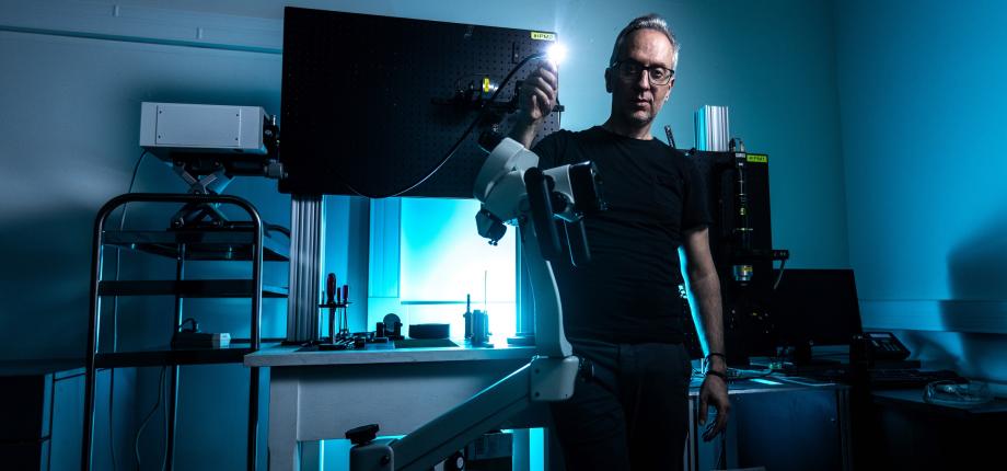
Angelo Pierangelo is a professor and researcher at École Polytechnique and has been leading research activities at LPICM on biophotonics since 2015. He has leading expertise in optical instrumentation, particularly in the field of Mueller polarimetric imaging for biomedical applications. His innovative approach is based on exploiting the properties of polarized light to decipher the complex microstructure of living tissues, paving the way for new strategies for the diagnosis and treatment of many diseases. A true driver of innovation, Angelo Pierangelo designs Mueller polarimetric imagers with unrivalled performance, and is one of the pioneers in the application of such systems in vivo. His flagship applications are numerous and have a direct clinical impact, including early detection of cervical cancer, improved diagnosis of prematurity, and the development of endoscopic systems for exploring internal organs and minimally invasive surgery. Angelo Pierangelo is the leader of the POLARIMA project, funded by the Fondation de l'X, which is a decisive step towards the implementation and deployment of his technology in a clinical setting. Angelo Pierangelo has extensive experience in managing complex multidisciplinary projects at the interface between physics, engineering, artificial intelligence, and the medical world. He has contributed significantly to the development of several collaborations with various research centers in physics and data processing (Xlim, ICUBE, Charles Fabry Laboratory, etc.) and hospitals (Institut Mutualiste Montsouris, Institut Gustave Roussy, Kremlin-Bicêtre University Hospital, Foch Hospital, etc.).
* LPICM : a joint research unit CNRS, École Polytechnique, Institut Polytechnique de Paris, 91120 Palaiseau, France
** CMAP : a joint research unit CNRS, Inria, École Polytechnique, Institut Polytechnique de Paris, 91120 Palaiseau, France